Cell Biology Explained: Analyze Diagrams on Your Screen with AI
Struggling with complex cell biology diagrams? Learn key structures, functions, and how AI screen assistants can help you study organelles and processes faster.
Try ScreenHelp Free
Get AI-powered screen assistance for any task. Analyze screenshots and get instant guidance.
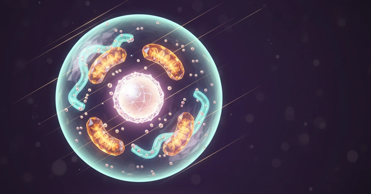
Get StartedCell biology is one of the most visual disciplines in all of science. From the intricate folds of the endoplasmic reticulum to the double-membrane structure of mitochondria, understanding cells requires you to see and interpret complex diagrams — not just memorize definitions.
Whether you're preparing for AP Biology, a college-level cell biology exam, or a medical certification, this guide breaks down the key concepts you'll encounter in cell diagrams and shows you how AI-powered screen analysis can accelerate your learning.
Why Cell Biology Diagrams Are So Challenging
Unlike some subjects where text-based studying is enough, cell biology demands visual literacy. A typical exam question might show you an electron micrograph or a labeled diagram and ask you to:
- Identify an organelle based on its structure
- Explain the function of a highlighted component
- Trace a pathway (like protein synthesis) across multiple organelles
- Compare prokaryotic and eukaryotic cell features
The challenge is that many organelles look similar at first glance, and their functions are deeply interconnected. Simply reading a textbook often isn't enough — you need to practice interpreting visual information.
Essential Cell Structures You Must Know
Let's review the core organelles and structures you'll encounter in virtually every cell biology course.
The Plasma Membrane
The phospholipid bilayer is the cell's gatekeeper. In diagrams, look for:
- Two parallel lines representing the hydrophilic heads and hydrophobic tails
- Embedded proteins — integral (spanning the membrane) and peripheral (attached to one side)
- Cholesterol molecules wedged between phospholipids, regulating fluidity
- Glycoproteins and glycolipids on the extracellular surface, involved in cell recognition
A common exam question involves the fluid mosaic model — understanding that the membrane is dynamic, not rigid.
The Nucleus
Often the most prominent structure in eukaryotic cell diagrams:
- Double membrane (nuclear envelope) with nuclear pores
- Nucleolus — a dense region where ribosomal RNA is synthesized
- Chromatin — loosely organized DNA (condensed into chromosomes during division)
Pay attention to the connection between the outer nuclear membrane and the rough endoplasmic reticulum — they're continuous.
Endoplasmic Reticulum (ER)
This is where many students get confused in diagrams:
- Rough ER — studded with ribosomes, appears bumpy; responsible for protein synthesis and modification
- Smooth ER — no ribosomes, appears tubular; involved in lipid synthesis, detoxification, and calcium storage
Tip: In diagrams, the rough ER is typically drawn closer to the nucleus, while smooth ER extends further into the cytoplasm.
Mitochondria
The "powerhouse of the cell" has distinctive features in diagrams:
- Outer membrane — smooth boundary
- Inner membrane — highly folded into cristae (this is the key visual identifier)
- Matrix — the interior space where the Krebs cycle occurs
- Intermembrane space — where the proton gradient forms during oxidative phosphorylation
Golgi Apparatus
Look for a stack of flattened, disc-like sacs (cisternae):
- Cis face — receiving side (near the ER)
- Trans face — shipping side (facing the plasma membrane)
- Transport vesicles budding off from the edges
The Golgi modifies, sorts, and packages proteins — think of it as the cell's post office.
Lysosomes and Peroxisomes
Both appear as small, circular, membrane-bound vesicles in diagrams:
- Lysosomes contain hydrolytic enzymes for intracellular digestion (pH ~5)
- Peroxisomes handle oxidative reactions and break down fatty acids
Distinguishing these visually can be tricky — exam diagrams usually label them or test your knowledge of their functions.
Cytoskeleton
Often overlooked in studying but frequently tested:
- Microfilaments (actin) — thinnest; involved in cell movement and shape
- Intermediate filaments — provide mechanical strength
- Microtubules — thickest; form the mitotic spindle, cilia, and flagella; composed of tubulin
Key Cellular Processes to Trace in Diagrams
Beyond identifying structures, exams often ask you to follow a process through the cell.
The Protein Synthesis Pathway
- Transcription in the nucleus → mRNA produced
- mRNA exits through nuclear pores
- Translation on ribosomes (free or on rough ER)
- Protein enters the rough ER lumen for folding
- Transport vesicle carries protein to the Golgi apparatus
- Golgi modifies and sorts → secretory vesicle
- Vesicle fuses with the plasma membrane (exocytosis)
Being able to trace this pathway on an unlabeled diagram is a common and high-value exam skill.
Cellular Respiration Overview
- Glycolysis — cytoplasm (doesn't require mitochondria)
- Pyruvate oxidation — mitochondrial matrix
- Krebs cycle — mitochondrial matrix
- Oxidative phosphorylation — inner mitochondrial membrane (cristae)
Photosynthesis (Plant Cells)
- Light reactions — thylakoid membranes inside chloroplasts
- Calvin cycle — stroma of the chloroplast
Prokaryotic vs. Eukaryotic Cells: A Visual Comparison
This is one of the most commonly tested diagram topics across all levels of biology.
| Feature | Prokaryotic | Eukaryotic |
|---|---|---|
| Nucleus | No (nucleoid region) | Yes (membrane-bound) |
| Membrane-bound organelles | No | Yes |
| Ribosomes | 70S | 80S |
| DNA structure | Circular, single | Linear, multiple chromosomes |
| Cell wall | Usually present (peptidoglycan in bacteria) | Sometimes (cellulose in plants, chitin in fungi) |
| Size | ~1-10 μm | ~10-100 μm |
When you encounter a diagram, the quickest way to determine if a cell is prokaryotic is the absence of a defined nucleus and membrane-bound organelles.
Plant vs. Animal Cells in Diagrams
Another frequent visual comparison:
- Cell wall — present in plant cells, absent in animal cells
- Chloroplasts — present in plant cells (and some protists)
- Central vacuole — large, dominant in plant cells; animal cells have smaller vacuoles
- Centrioles — typically in animal cells (used in cell division)
How AI Screen Analysis Helps You Study Cell Biology
Here's where modern study tools can make a real difference. When you're working through a textbook, an online lecture, or a practice exam and encounter a complex cell diagram, you don't always have time to cross-reference every structure manually.
An AI screen assistant like ScreenHelp lets you capture any diagram displayed on your screen and get an instant, detailed explanation. Here's what makes this approach effective for cell biology:
- Identify unlabeled structures: Share your screen, capture a diagram, and ask the AI to identify all visible organelles
- Understand relationships: Ask why certain organelles are positioned near each other or how they interact in a process
- Practice with custom prompts: Set up prompts like "Explain all structures in this cell diagram" or "Trace the protein synthesis pathway shown" so you can quickly analyze multiple diagrams during a study session
- Study anywhere: Use the QR code feature to stream AI explanations to your phone while working through a desktop textbook or lecture recording
Because ScreenHelp uses AI with vision capabilities, it can actually see and interpret the visual content on your screen — not just text, but the shapes, labels, arrows, and spatial relationships in biological diagrams.
Study Tips for Mastering Cell Biology Diagrams
1. Draw From Memory
After studying a diagram, close your materials and sketch the cell from memory. Label everything you can, then compare. The gaps in your drawing reveal exactly what you need to review.
2. Use Active Recall with Diagrams
Don't just stare at labeled diagrams. Cover the labels and quiz yourself. This is where an AI screen assistant becomes particularly useful — capture your unlabeled diagram and check your answers against the AI's analysis.
3. Connect Structure to Function
For every organelle, ask yourself: Why does it look this way? Mitochondria have folded inner membranes because more surface area means more ATP production. The rough ER has ribosomes because it synthesizes membrane-bound and secreted proteins. Structure always serves function.
4. Compare Across Cell Types
Practice with diagrams from different cell types — nerve cells, red blood cells, muscle cells, plant mesophyll cells. Notice how organelle proportions change based on cell function (e.g., muscle cells have abundant mitochondria).
5. Work Through Process Diagrams
Don't just memorize static cell diagrams. Practice with dynamic ones that show processes like mitosis, meiosis, endocytosis, or signal transduction. These test deeper understanding.
Common Mistakes on Cell Biology Exams
- Confusing smooth and rough ER: Remember, ribosomes on the rough ER are the distinguishing feature
- Forgetting the double membrane: Both the nucleus and mitochondria have double membranes — this connects to the endosymbiotic theory
- Ignoring scale: Ribosomes are far smaller than mitochondria; diagram proportions aren't always accurate
- Mixing up cilia and flagella function: Both involve microtubules, but cilia are short and numerous, flagella are long and few
- Overlooking the cytoskeleton: It's not just structural support — it's essential for transport, division, and cell movement
Final Thoughts
Cell biology is fundamentally a visual science. The better you get at reading and interpreting diagrams, the more confident you'll be on exams — from introductory biology to medical boards. Combine traditional study techniques like active recall and sketching with modern tools like AI screen analysis, and you'll build the kind of deep understanding that lasts well beyond test day.
Start Using AI Screen Assistance Today
Join thousands of users who are already working smarter with ScreenHelp.
Related Articles

Identifying Biological Structures in Lab Reports with AI
Struggling to identify cells, tissues, and organelles for lab reports? Learn how AI screen assistants can help you study biological structures more effectively.
Read article
The Ultimate Study Hack: Turning Your Screen into a Tutor
Discover how to transform your everyday screen into a powerful study companion using AI screen assistants that explain, clarify, and help you learn faster.
Read article
Translate and Understand Foreign Language Text on Screen Instantly
Struggling with foreign language text during studies? Learn how AI screen assistants can translate, explain grammar, and help you understand any language right on your screen.
Read article