Identifying Biological Structures in Lab Reports with AI
Struggling to identify cells, tissues, and organelles for lab reports? Learn how AI screen assistants can help you study biological structures more effectively.

Try ScreenHelp Free
Get AI-powered screen assistance for any task. Analyze screenshots and get instant guidance.
Get StartedThe Challenge of Biological Structure Identification
Every biology student knows the feeling: staring at a microscope slide image or a histology diagram, trying to distinguish between smooth endoplasmic reticulum and rough endoplasmic reticulum, or telling apart simple squamous epithelium from stratified columnar epithelium. Identifying biological structures is a foundational skill across biology, anatomy, histology, microbiology, and dozens of other life science courses — and it's one of the hardest to master.
Whether you're working on a formal lab report, preparing for a practical exam, or reviewing virtual slides, accurate identification is non-negotiable. A mislabeled organelle or misidentified tissue type doesn't just cost you points — it signals a gap in understanding that can compound as coursework advances.
So how can students get better at this essential skill? And how are modern AI tools changing the way students study visual biological content?
Why Biological Identification Is So Difficult
Visual Similarity Between Structures
Many biological structures look strikingly similar, especially under certain magnifications or staining techniques. Consider these commonly confused pairs:
- Mitochondria vs. chloroplasts in electron micrographs with ambiguous staining
- Smooth muscle vs. dense regular connective tissue in longitudinal sections
- Prokaryotic vs. eukaryotic cells at low magnification
- Arterioles vs. venules in cross-section without clear labeling
Without extensive practice, even diligent students mix these up regularly.
Variability in Specimen Preparation
The same tissue can look dramatically different depending on the staining method (H&E vs. PAS vs. Masson's trichrome), section angle (transverse vs. longitudinal vs. oblique), and specimen quality. A real lab specimen rarely looks as clean as a textbook diagram, which makes transferring knowledge from study materials to actual lab work surprisingly difficult.
Limited Feedback Loops
In a traditional lab setting, you might get feedback once — when the graded report comes back. That delay means you could practice incorrectly for days before learning about a misidentification. Faster feedback dramatically improves learning outcomes in visual identification tasks.
Strategies for Better Biological Structure Identification
Before discussing AI tools, let's cover proven study strategies that every life science student should use.
1. Build a Personal Visual Atlas
As you encounter structures in lectures, labs, and textbooks, compile your own collection of annotated images. Organizing them by organ system, tissue type, or staining method creates a personalized reference that mirrors the way you actually learn.
2. Use the "Describe Before You Label" Method
Before naming a structure, force yourself to describe its visual characteristics objectively:
- What is the shape?
- What is the relative size compared to surrounding structures?
- What color does it stain?
- Where is it located in relation to other identifiable landmarks?
This builds diagnostic reasoning skills rather than pattern-matching from memorization alone.
3. Study with Unlabeled Images
Seek out unlabeled versions of diagrams and micrographs. Many histology atlases and online resources offer practice images without annotations. Test yourself, then check against labeled versions.
4. Learn Staining Logic
Understanding why structures stain certain colors (e.g., H&E stains nuclei blue-purple because hematoxylin binds to acidic DNA) gives you a deductive framework instead of requiring rote memorization of every possible appearance.
5. Form Study Groups for Practical Review
Verbalizing identifications to peers — and hearing their reasoning — exposes gaps in your understanding that solo study often misses.
How AI Vision Tools Are Changing the Game
Recent advances in AI, particularly in large language models with vision capabilities, have opened a new avenue for studying biological structures. These tools can analyze images on your screen and provide detailed explanations of what they see.
Instant Explanations of Visual Content
Imagine you're reviewing a histology slide on your computer and you're unsure whether you're looking at stratified squamous epithelium or transitional epithelium. An AI screen assistant can analyze what's displayed on your screen and walk you through the distinguishing features — the cell layer arrangement, the shape of surface cells, the presence or absence of a basement membrane — giving you an on-demand explanation.
This kind of instant, contextual feedback is something that was previously only available during office hours or with a tutor sitting beside you.
Learning the Reasoning, Not Just the Answer
The best way to use AI for biological identification isn't just asking "What is this?" It's asking the AI to explain its reasoning. For example:
- "What features in this image indicate this is cardiac muscle rather than skeletal muscle?"
- "Walk me through how to differentiate these two tissue types based on what's visible."
- "What staining characteristics should I look for to confirm this is a goblet cell?"
This approach turns the tool into a tutor that teaches you the diagnostic process, which is far more valuable than simple identification.
Working with Virtual Labs and Digital Slides
Many universities now use virtual microscopy platforms (like Aperio, PathPresenter, or university-hosted whole slide imaging systems). Since these are viewed on-screen, they're a natural fit for AI screen assistants. You can study digital slides at your own pace while getting AI-powered explanations of the structures you're examining.
Using ScreenHelp for Biology Study Sessions
ScreenHelp is an AI screen assistant that can see what's on your display and provide intelligent responses. For biology students, it works well as a study companion for visual identification tasks.
Here's how the workflow typically looks:
- Open your study material — a virtual slide, digital atlas, lab manual PDF, or lecture recording with microscopy images
- Start ScreenHelp by clicking the browser button, which initiates a screen share
- Trigger the AI when you encounter a structure you want to understand better
- Read the explanation directly in your browser, or scan the QR code to stream the response to your phone (handy if your computer screen is filled with the specimen image)
You can also set up custom predefined prompts tailored to biology study, such as:
- "Identify all visible biological structures in this image and explain their functions"
- "What tissue type is shown? Describe the key identifying features"
- "List the organelles visible in this electron micrograph and their roles"
- "Compare the structures visible here to what I would see with a different staining method"
With the browser extension, you can trigger captures using keyboard shortcuts from anywhere on your computer — useful when working in dedicated virtual microscopy software outside the browser.
Specific Use Cases in the Life Sciences
Histology
Histology is perhaps the most natural fit for AI-assisted visual learning. The discipline is entirely about identifying tissues from visual characteristics. Students can use AI to verify identifications of the four basic tissue types (epithelial, connective, muscle, and nervous) and their many subtypes.
Cell Biology
Electron micrographs of cellular organelles can be analyzed for structure identification. AI can help explain the visual differences between organelles like the Golgi apparatus, lysosomes, and peroxisomes, which can appear similar in TEM images.
Microbiology
Bacterial morphology (cocci, bacilli, spirilla), Gram stain results, and colony characteristics on culture plates are all visual identification tasks where AI assistance can accelerate learning.
Anatomy
Whether studying cross-sectional anatomy on CT/MRI images or cadaver photographs, AI can help identify anatomical structures and describe their spatial relationships.
Botany
Plant tissue identification — distinguishing xylem from phloem, identifying meristematic vs. permanent tissues, or recognizing different leaf anatomies — follows the same visual identification pattern.
Tips for Getting the Most Out of AI-Assisted Biology Study
-
Always verify against authoritative sources. AI is a study aid, not a replacement for your textbook or instructor. Cross-reference identifications with your course materials.
-
Ask for reasoning, not just labels. The learning happens when you understand why a structure is identified a certain way.
-
Use it as a first pass, then test yourself. Study a set of structures with AI help, then come back later and try to identify them on your own.
-
Provide context in your prompts. Telling the AI what staining method was used, what organ the sample is from, or what magnification you're viewing at will improve the accuracy of its analysis.
-
Don't skip the fundamentals. AI tools are most effective when you already have a foundation. They help you fill gaps and refine knowledge — they're less effective as a complete substitute for attending lectures and reading the material.
The Future of Visual Learning in Biology
As AI vision models continue to improve, the accuracy and depth of real-time biological structure identification will only get better. For now, tools like ScreenHelp offer a practical way to get instant feedback during study sessions — something that can meaningfully accelerate the learning curve for visual identification skills.
The students who will benefit most are those who use these tools actively and critically: questioning the AI's reasoning, comparing its answers with textbook descriptions, and using the feedback loop to genuinely improve their own identification abilities.
Start Using AI Screen Assistance Today
Join thousands of users who are already working smarter with ScreenHelp.
Related Articles
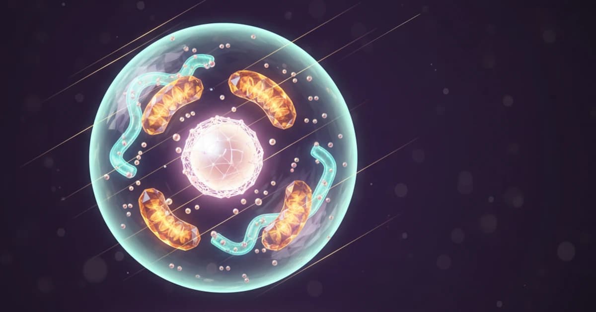
Cell Biology Explained: Analyze Diagrams on Your Screen with AI
Struggling with complex cell biology diagrams? Learn key structures, functions, and how AI screen assistants can help you study organelles and processes faster.
Read article
Translate and Understand Foreign Language Text on Screen Instantly
Struggling with foreign language text during studies? Learn how AI screen assistants can translate, explain grammar, and help you understand any language right on your screen.
Read article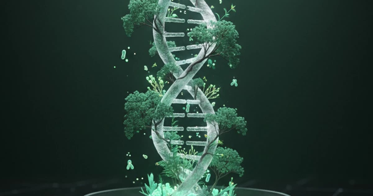
From DNA to Ecology: The AI Tool for Biology Students
Biology covers vast topics from molecular genetics to ecosystems. Discover how AI screen assistants help biology students study smarter and master complex concepts.
Read article